Introduction
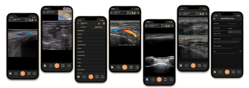
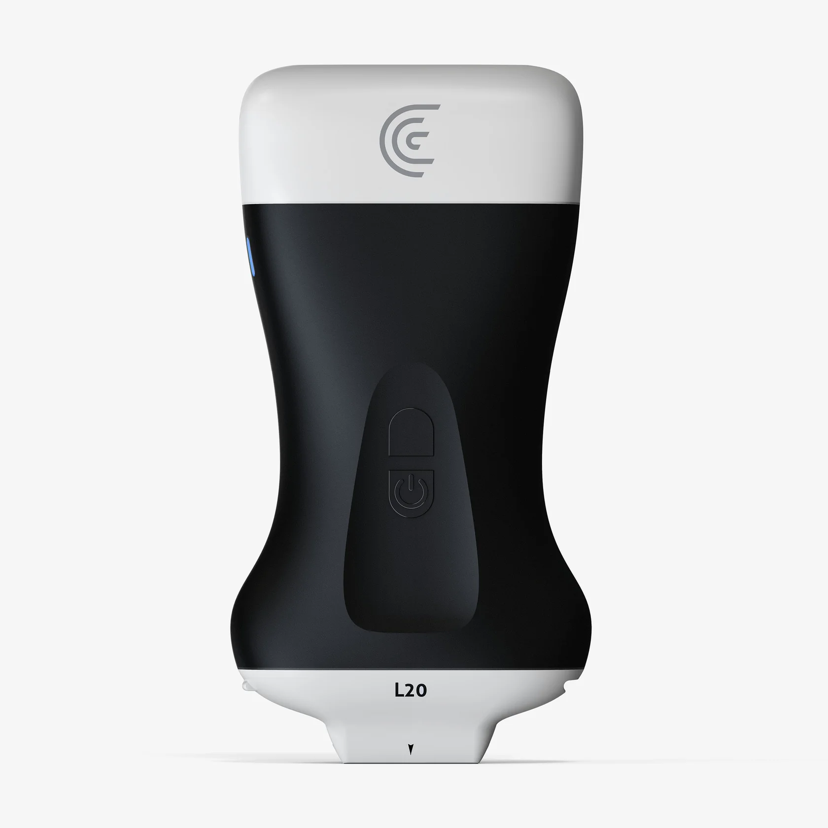
The Clarius L20 ultrasound scanner is a high-frequency, wireless device designed to provide superior imaging of superficial structures. It is particularly suited for aesthetic applications such as dermal fillers, facial rejuvenation, and vascular mapping. This comprehensive guide offers detailed instructions on setting up and optimising the use of the Clarius L20 scanner, including ideal initial settings, optimal use of various modes, and in-depth information on the sonographic appearance of different tissues relevant to aesthetic practice.

1. Setting Up the Clarius L20 Scanner
1.1. Unboxing and Charging
- Careful Unpacking:
- Remove the scanner, battery, charging station, and accessories from the packaging with care.
- Battery Charging:
- Insert the battery into the scanner securely.
- Place the scanner on the charging station.
- Charge until the battery indicator shows full capacity (approximately 90 minutes).
1.2. Installing the Clarius App
- App Download:
- For iOS devices, visit the App Store.
- For Android devices, access the Google Play Store.
- Search for “Clarius Ultrasound” and install the application.
- Account Creation:
- Open the app and create a Clarius Cloud account, or log in if you already possess one.
- Permission Settings:
- Allow necessary permissions for Wi-Fi, Bluetooth, and location services to ensure optimal connectivity.
1.3. Connecting the Scanner to Your Device
- Powering On:
- Press and hold the power button on the scanner until the LED indicator lights up.
- Wi-Fi Connection:
- The scanner creates its own Wi-Fi network.
- Navigate to your device’s Wi-Fi settings and connect to the network named “Clarius-XXXX”.
- App Integration:
- Open the Clarius app.
- The scanner should connect automatically.
- If prompted, update the scanner firmware to the latest version for optimal performance.
2. Ideal Initial Settings for Aesthetic Use
2.1. Preset Selection
- Appropriate Preset Choice:
- Select the “Superficial” or “Cosmetic” preset if available.
- These presets are optimised for high-resolution imaging of superficial tissues.
2.2. Frequency Settings
- High-Frequency Imaging:
- Set the frequency to the highest available (15–20 MHz) to achieve maximum resolution.
- Ideal for visualising superficial structures such as skin layers and small vessels.
2.3. Depth Adjustment
- Optimal Depth Setting:
- Adjust the depth to 1–3 cm to focus on superficial tissues.
- This enhances image quality by concentrating ultrasound energy where needed.
2.4. Gain and Time Gain Compensation (TGC)
- Gain Adjustment:
- Begin with a medium gain setting.
- Incrementally adjust to optimise image brightness and contrast.
- TGC Sliders:
- Fine-tune the brightness at different depths to ensure uniform image quality from superficial to deeper structures.
2.5. Focus Position
- Focal Zone Placement:
- Position the focal zone at or just below the area of interest.
- Enhances image resolution in the region requiring the most detail.
2.6. Image Optimisation Settings
- Dynamic Range:
- Set to a medium level to balance contrast and detail visibility.
- Persistence:
- Low to medium settings reduce noise without blurring moving structures.
- Speckle Reduction:
- Utilise this feature to minimise speckle artefacts and improve image clarity.
3. Optimal Use of Various Modes
3.1. B-Mode (2D Mode)
- Primary Imaging Mode:
- Use for general visualisation of anatomical structures.
- Optimisation Tips:
- Adjust gain and depth settings for optimal image clarity.
- Employ freeze and zoom functions to examine specific areas in detail.
3.2. Colour Doppler Mode
- Vascular Mapping:
- Activate to visualise blood flow within vessels.
- Optimisation Tips:
- Colour Gain:
- Adjust to a level where colour fills the vessel lumen without extending beyond its walls.
- Pulse Repetition Frequency (PRF):
- Set low for detecting slow-flow vessels common in superficial tissues.
- Wall Filter:
- Adjust to eliminate low-frequency noise while preserving true flow signals.
- Steering Angle:
- Align the Doppler beam with the direction of blood flow for enhanced detection.
3.3. Power Doppler Mode
- Sensitive Flow Detection:
- More sensitive than Colour Doppler for detecting low-velocity flow.
- Optimisation Tips:
- Adjust settings to maximise sensitivity without introducing excessive noise.
- Ideal for detecting microvascular flow in superficial tissues.
3.4. M-Mode
- Motion Analysis:
- Not commonly used in aesthetics but can assess moving structures if required.
- Optimisation Tips:
- Adjust sweep speed to capture the motion of interest accurately.
4. Performing the Ultrasound Examination
4.1. Patient Preparation
- Positioning:
- Ensure the patient is comfortably positioned with the area of interest easily accessible.
- Skin Preparation:
- Clean the skin thoroughly to remove any debris or makeup.
- Gel Application:
- Apply a small amount of ultrasound gel to the area or directly onto the probe to facilitate acoustic coupling.
4.2. Scanning Technique
- Probe Handling:
- Hold the probe lightly to avoid excessive pressure that could compress superficial vessels or distort tissues.
- Orientation Awareness:
- Be mindful of the probe marker and screen orientation to interpret images correctly.
- Systematic Scanning:
- Scan in both longitudinal and transverse planes for a comprehensive assessment.
4.3. Identifying Anatomical Structures
- Layer Visualisation:
- Identify the various skin layers, subcutaneous tissue, muscles, and vessels.
- Vessel Identification:
- Use Colour or Power Doppler to locate arteries and veins accurately.
- Anomaly Detection:
- Be vigilant for any unexpected structures or pathologies that may impact treatment.
5. Ultrasound-Guided Procedures
5.1. Needle Guidance
- Technique Selection:
- In-Plane Approach:
- The needle is aligned with the ultrasound beam, providing full visualisation of the needle shaft and tip.
- Out-of-Plane Approach:
- The needle crosses the ultrasound beam, appearing as a cross-section on the image.
- Needle Visualisation:
- Use high-frequency settings to enhance needle clarity.
- Adjust the angle of insertion to reduce artefacts and improve visibility.
5.2. Safety Considerations
- Avoiding Vessels:
- Employ Doppler modes to identify and steer clear of blood vessels during injections.
- Real-Time Monitoring:
- Continuously observe needle placement and filler distribution to ensure accuracy.
- Patient Communication:
- Keep the patient informed about the procedure to minimise anxiety and encourage cooperation.
6. Post-Examination Procedures
6.1. Image Saving and Documentation
- Capturing Images:
- Use the app to save still images or video loops of significant findings.
- Annotations:
- Add labels, measurements, or notes directly onto the images for future reference.
- Data Storage:
- Securely save images to the Clarius Cloud or export them to your electronic medical records (EMR) system.
6.2. Cleaning and Maintenance
- Probe Cleaning:
- Wipe the probe with approved disinfectant wipes, following manufacturer guidelines.
- Avoiding Damage:
- Ensure no liquids enter the battery compartment or connectors.
- Proper Storage:
- Place the scanner back on the charging station or in its protective case when not in use.
7. Safety Precautions and Contraindications
7.1. Patient Safety
- Medical History Review:
- Assess the patient’s medical history for any contraindications to ultrasound use or aesthetic procedures.
- Skin Integrity:
- Avoid scanning over areas with compromised skin unless clinically necessary.
- Allergy Checks:
- Confirm that the patient has no known allergies to ultrasound gel or procedural materials.
- Patient Comfort:
- Ensure the patient is comfortable and informed throughout the procedure.
7.2. Operator Safety
- Ergonomics:
- Maintain proper posture and hand positioning to prevent strain during scanning.
- Infection Control:
- Adhere to standard infection control protocols, including hand hygiene and use of personal protective equipment (PPE).
- Electrical Safety:
- Regularly inspect the scanner and accessories for damage that could pose hazards.
7.3. Equipment Care
- Regular Maintenance:
- Follow the manufacturer’s maintenance schedule to ensure optimal performance.
- Software Updates:
- Keep the scanner’s firmware and app software up to date.
7.4. Contraindications
- Absolute Contraindications:
- Do not use ultrasound over areas with known malignant tumours unless specifically indicated.
- Relative Contraindications:
- Exercise caution when scanning patients with electronic implants, consulting device-specific guidelines.
8. Compliance with Regulatory Standards
8.1. Data Protection and Privacy
- Confidentiality:
- Comply with the General Data Protection Regulation (GDPR) by ensuring patient data is securely stored and transmitted.
- Consent:
- Obtain informed consent for both the ultrasound examination and any data storage or sharing.
- Anonymisation:
- Remove all patient identifiers when sharing images for educational purposes.
8.2. Professional Standards
- Qualifications:
- Ensure you are adequately trained and certified to perform ultrasound examinations in aesthetic practice.
- Clinical Governance:
- Adhere to guidelines from professional bodies such as the British Medical Ultrasound Society (BMUS).
- Documentation:
- Maintain accurate records of all procedures in accordance with legal requirements.
9. Enhancing Image Interpretation Skills
9.1. Understanding Ultrasound Physics
- Acoustic Properties:
- Familiarise yourself with how different tissues reflect ultrasound waves.
- Artefact Recognition:
- Learn to identify common artefacts such as shadowing, enhancement, and reverberation.
9.2. Anatomical Knowledge
- Superficial Anatomy:
- Deepen your understanding of facial anatomy, including skin layers, fat pads, muscles, and vasculature.
- Anatomical Variations:
- Be aware of variations that may affect procedural planning.
9.3. Continuing Professional Development
- Workshops and Courses:
- Attend advanced training sessions to stay updated on the latest techniques.
- Peer Collaboration:
- Engage with colleagues to discuss cases and share insights.
10. Advanced Imaging Techniques
10.1. Elastography (if available)
- Tissue Characterisation:
- Use elastography to assess tissue stiffness, aiding in the identification of fibrosis or scarring.
- Optimisation Tips:
- Apply consistent pressure during scanning for accurate results.
10.2. Three-Dimensional Imaging (if available)
- Volumetric Analysis:
- Utilise 3D imaging to assess volume changes post-procedure.
- Patient Communication:
- Use 3D images to help patients visualise treatment plans and outcomes.
11. Incorporating Ultrasound into Aesthetic Practice
11.1. Pre-Procedural Planning
- Assessment:
- Use ultrasound to evaluate the treatment area, identifying vital structures and planning injection sites.
- Customisation:
- Tailor treatments based on individual anatomical findings.
11.2. Post-Procedural Evaluation
- Monitoring:
- Assess the immediate effects of procedures, ensuring correct placement of fillers.
- Complication Management:
- Identify and address complications such as vascular occlusion promptly using ultrasound guidance.
11.3. Patient Education
- Visual Aids:
- Show patients ultrasound images to explain procedures.
- Trust Building:
- Demonstrating thoroughness through ultrasound use can enhance patient confidence.
12. Ethical Considerations
12.1. Scope of Practice
- Competency:
- Only perform procedures for which you are trained and qualified.
- Referral:
- Refer patients to specialists when findings are beyond your scope.
12.2. Patient Autonomy
- Informed Decisions:
- Provide all necessary information for patients to make informed choices.
- Right to Decline:
- Respect a patient’s decision to refuse any part of the procedure.
13. Detailed Sonographic Appearance of Tissues
Understanding the ultrasound characteristics of various anatomical structures is crucial for accurate interpretation and effective application in aesthetic practice. Below is an in-depth analysis of how different tissues and materials appear on ultrasound imaging using the Clarius L20 scanner.
13.1. Skin Layers
Epidermis and Dermis
- Appearance:
- Epidermis:
- Appears as a thin, superficial echogenic line due to its dense cellular composition.
- Dermis:
- Located just below the epidermis, presenting as a slightly hypoechoic band with echogenic strands representing collagen fibres.
- Tips for Identification:
- Use high-frequency settings (15–20 MHz) to enhance resolution.
- Adjust gain to prevent over-brightening, which can obscure details.
13.2. Subcutaneous Fat (Hypodermis)
- Appearance:
- Presents as a hypoechoic layer with echogenic linear septations representing connective tissue.
- Fat lobules appear as dark areas separated by bright connective tissue strands.
- Tips for Identification:
- Moderate gain settings help differentiate fat lobules from surrounding tissues.
- Avoid excessive pressure to prevent compressing the fat layer.
13.3. Muscles
- Appearance:
- Longitudinal View:
- Display a striated pattern of parallel hypoechoic muscle fibres separated by echogenic lines.
- Transverse View:
- Exhibit a “starry night” appearance with hypoechoic muscle fibres interspersed with echogenic connective tissue.
- Tips for Identification:
- Dynamic scanning (asking the patient to contract the muscle) can help confirm muscle tissue.
- Adjust the depth to include the entire muscle thickness.
13.4. Bones
- Appearance:
- Highly echogenic and appear as bright, continuous lines with posterior acoustic shadowing.
- Tips for Identification:
- Recognise the sharp interface and shadowing to differentiate bone from other echogenic structures.
- Bones serve as important landmarks for depth assessment during procedures.
13.5. Ligaments and Tendons
- Appearance:
- Both are highly echogenic due to dense collagen content.
- Tendons:
- Show a fibrillar pattern with parallel echogenic lines in the longitudinal view.
- Ligaments:
- Similar to tendons but may be shorter and connect bone to bone.
- Tips for Identification:
- Use the highest frequency setting for maximum resolution.
- Apply minimal pressure to avoid compressing and distorting the structures.
13.6. Blood Vessels
Arteries
- Appearance:
- Anechoic tubular structures with pulsatile flow.
- Vessel walls may be visible as echogenic outlines.
- Doppler Characteristics:
- Colour Doppler:
- Shows pulsatile, high-velocity flow with colour filling the lumen.
- Tips for Identification:
- Use Colour Doppler to confirm pulsatility and flow direction.
- Adjust the Doppler angle for optimal flow detection.
Veins
- Appearance:
- Anechoic tubular structures with thinner walls.
- Compressible and may show spontaneous low-velocity flow.
- Doppler Characteristics:
- Colour Doppler:
- Displays continuous, non-pulsatile flow.
- Tips for Identification:
- Apply gentle pressure to assess compressibility.
- Use Power Doppler to detect low-velocity venous flow.
13.7. Nerves
- Appearance:
- Transverse View:
- Display a “honeycomb” pattern with multiple hypoechoic fascicles surrounded by echogenic connective tissue.
- Longitudinal View:
- Show parallel hypoechoic bands separated by echogenic lines.
- Tips for Identification:
- Use both views to confirm nerve identification.
- Be cautious to avoid nerve injury during procedures.
13.8. Previous Dermal Fillers
Hyaluronic Acid Fillers
- Appearance:
- Typically hypoechoic to anechoic areas within the soft tissue.
- May appear well-defined or diffuse depending on injection technique and time since placement.
- Tips for Identification:
- Recent fillers may appear more anechoic due to higher water content.
- Use dynamic scanning to differentiate filler material from cysts or other fluid collections.
Calcium Hydroxylapatite Fillers
- Appearance:
- Echogenic foci with posterior acoustic shadowing due to calcium particles.
- Tips for Identification:
- Recognise the shadowing effect to differentiate from other echogenic structures.
- Adjust gain and frequency settings for optimal visualisation.
Poly-L-Lactic Acid Fillers
- Appearance:
- May not be readily visible initially.
- Over time, can induce echogenic fibrous tissue formation.
- Tips for Identification:
- Look for subtle changes in tissue echogenicity.
- Correlate with the patient’s treatment history.
13.9. Scar Tissue and Fibrosis
- Appearance:
- Irregular, echogenic areas within the soft tissue.
- Tips for Identification:
- Use a combination of B-Mode and elastography to assess fibrosis extent.
- Differentiate from normal connective tissue by the irregular pattern.
13.10. Cysts and Fluid Collections
- Appearance:
- Anechoic areas with well-defined borders.
- May exhibit posterior acoustic enhancement.
- Tips for Identification:
- Use both longitudinal and transverse scans.
- Differentiate from solid masses by the lack of internal echoes.
13.11. Foreign Bodies
- Appearance:
- Metallic Objects:
- Highly echogenic with significant shadowing.
- Organic Materials:
- May appear hyperechoic with posterior shadowing or enhancement.
- Tips for Identification:
- Adjust frequency and gain to improve contrast.
- Consider the patient’s history of exposure.
14. Practical Application in Aesthetic Procedures
14.1. Assessment of Injection Depth
- Technique:
- Use echogenic layers as a guide for appropriate filler placement depth.
- Outcome:
- Avoid over-injection and monitor tissue expansion in real-time.
14.2. Avoiding Vital Structures
- Technique:
- Identify and map out blood vessels and nerves prior to injections.
- Outcome:
- Adjust needle trajectory based on anatomical findings to enhance safety.
14.3. Monitoring Filler Distribution
- Technique:
- Observe filler dispersion during injection.
- Outcome:
- Ensure even distribution and correct irregularities immediately.
15. Enhancing Ultrasound Imaging Skills
15.1. Mastering Equipment Settings
- Frequency Selection:
- Use the highest frequency that allows adequate penetration for target tissue depth.
- Optimising Gain and Focus:
- Adjust gain to enhance contrast without introducing noise.
- Set the focal zone at the level of interest.
15.2. Image Interpretation Techniques
- Systematic Approach:
- Scan in multiple planes and compare bilateral structures.
- Artefact Recognition:
- Identify and understand common artefacts to avoid misinterpretation.
15.3. Continuous Learning
- Stay Updated:
- Engage with current literature and attend workshops.
- Peer Collaboration:
- Share experiences with colleagues to enhance collective knowledge.
16. Common Artefacts and How to Recognise Them
16.1. Acoustic Shadowing
- Description:
- Dark areas beneath echogenic structures like bone.
- Recognition:
- Acknowledge that structures distal to the shadow may not be visible.
16.2. Posterior Acoustic Enhancement
- Description:
- Increased brightness beneath anechoic structures like cysts.
- Recognition:
- Useful in confirming fluid-filled lesions.
16.3. Reverberation Artefacts
- Description:
- Multiple, equally spaced echogenic lines.
- Recognition:
- Often seen with metallic objects or air bubbles.
16.4. Mirror Image Artefact
- Description:
- Duplication of structures due to reflection off strong interfaces.
- Recognition:
- Avoid misinterpreting these as actual anatomical structures.
17. Conclusion and Best Practices
Integrating the Clarius L20 ultrasound scanner into aesthetic practice elevates patient care by enhancing precision and safety. Adherence to optimal settings and proficient use of imaging modes allows for detailed visualisation of superficial structures, facilitating better clinical outcomes.
Best Practices Summary
- Preparation:
- Ensure both equipment and patient are adequately prepared.
- Optimisation:
- Continuously adjust settings for the best image quality.
- Education:
- Invest in ongoing training to stay abreast of advancements.
- Safety:
- Prioritise safety through adherence to protocols and guidelines.
- Professionalism:
- Maintain ethical standards and comply with all regulatory requirements.
References
- Adembri, C., et al. (2013). “Ultrasound in aesthetic medicine: vascular mapping to avoid complications.” Journal of Cosmetic Dermatology, 12(2), 155–161.
- Bianchi, S., & Martinoli, C. (2007). Ultrasound of the Musculoskeletal System. Springer.
- Huang, Y. C., et al. (2013). “Sonographic features of keloids and hypertrophic scars.” Journal of Medical Ultrasound, 21(1), 20–24.
- Kapoor, K. M., et al. (2020). “Use of ultrasound in the safe delivery of fillers in the face.” Journal of Cutaneous and Aesthetic Surgery, 13(2), 142–148.
- Tagliafico, A., et al. (2010). “High-resolution ultrasound of the face and body.” American Journal of Roentgenology, 194(1), W6–W12.
- Urdiales-Gálvez, F., et al. (2017). “Treatment of soft tissue filler complications: expert consensus recommendations.” Aesthetic Plastic Surgery, 41(3), 667–677.
- Wortsman, X. (2012). “Sonography of cosmetic dermatologic complications.” Ultrasound Clinics, 7(3), 327–341.
- Wortsman, X. (2015). “Ultrasound in dermatology: why, how, and when?” Seminars in Ultrasound, CT and MRI, 36(3), 177–195.
Note: This comprehensive guide is intended to support practitioners in the aesthetic field by providing detailed, evidence-based information on the use of the Clarius L20 ultrasound scanner. Always refer to the latest user manual and professional guidelines provided by Clarius and relevant medical authorities. Ensure compliance with local regulations and engage in continuous professional development to maintain the highest standards of patient care.


